

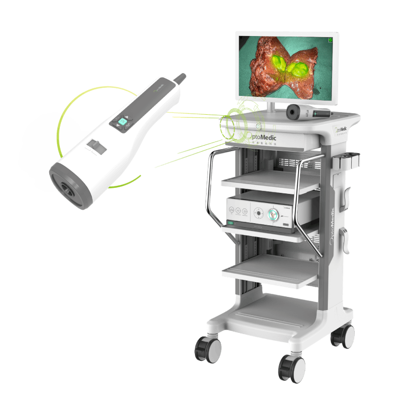
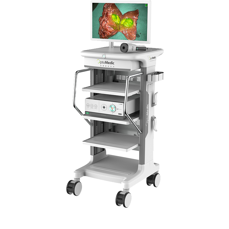
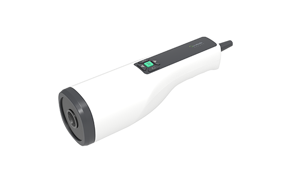
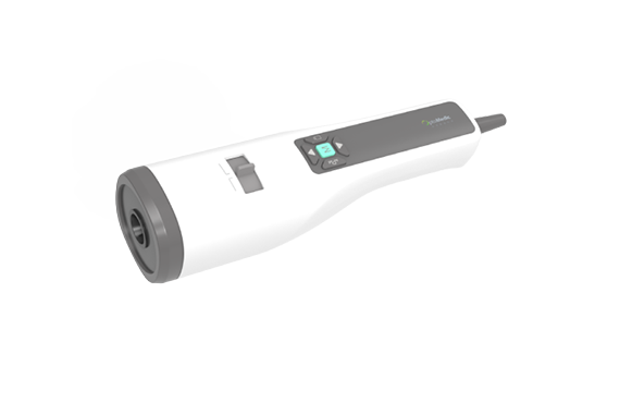
The FloNavi Open Field Fluorescence Imaging System is an imaging system used in hospitals for capturing and viewing fluorescent images for the visual assessment of blood flow, as an adjunctive method for the evaluation of tissue perfusion, and related tissue transfer circulation for use in imaging during various surgical procedures. The FloNavi Open Field Fluorescence Imaging System includes an Imaging Head, an Image Processing Unit, and power supply cords. The Imaging Head may be either hand-held or attached to a mechanical arm and provides illumination of the regions of a patient\'s body to be observed with near infrared light to excite ICG fluorescence. Alternatively, the Imaging Head provides white light illumination of the regions of a patient\'s body to be observed for color imaging. The cameras in the Imaging Head capture the fluorescent image under near infrared illumination or a color image under white light illumination. The Image Processing Unit receives the video signal from the Imaging Head and processes and outputs the video image to a medical grade video monitor and/or video recorder. Adjustments to the operation of the FloNavi Open Field Fluorescence Imaging System are possible through switches at either the Imaging Head or the Image Processing Unit.
ICG, Indocyanine Green Dye, featured with no side effects, wide application, and low cost, has been clinically approved for many years. It enters the human lymph and blood circulation through tumor surrounding tissue or intravenous injection.
ICG will bind the plasma lipoproteins.
After exposed to NIR with a wavelength of 805 nm, proteins will emit fluorescence signal with a wavelength of 835 nm.
NOTE:
ICG contains sodium iodide and should be used with caution in patients who have a history of allergy to iodides or iodinated imaging agents due to a risk of anaphylaxis.
The Imaging System should not be used for NIR imaging during surgical procedures with patients who are known to be sensitive to iodides or iodinated imaging agents.
Radioactive iodine uptake studies should not be performed for at least a week following the use of ICG for injection.
Safety and Performance Information
Brilliant High-definition Imaging
The Precision Fluorescence Imaging System
Various Fluorescence Imaging Modes for Open Surgeries
|
Model |
OPTO-CHD3100E, OPTO-CHD3100H |
|
Manufacturer |
Guangdong OptoMedic Technologies, Inc. |
|
Protection against electric shock Class 1 |
|
|
Dimensions (L×H×W) |
250 mm×77 mm×77 mm |
|
Cable length |
3 m |
|
Image resolution |
1920×1080p |
|
Video resolution |
1920×1080p, 720×576i, when used with the FloNavi Image Process Unit |
|
Near infrared LED array |
Wavelength: 785 nm±10 nm |
|
Lamp classification |
Risk group 0 (as per IEC/TR 62471-2) |
|
Optical window cover plate |
Model: CS690 |
|
Compatible control unit |
FloNavi Image Processing Unit (OPTO-CAM2100) |
|
Electrical power |
Refer to the operator’s manual of compatible control unit |


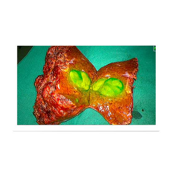
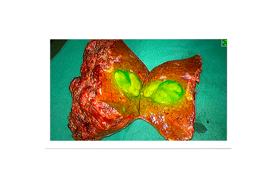
Sentinel lymph node tracing of breast cancer

Autofluorescence for parathyroid gland
Lymph node mapping
Gland perfusion

Liver segments visualization - Anatomical liver segmentectomy
Tumor Identification - Accurate display of tumor location, margin, and microsatellite
Biliary tract visualization - Complex biliary tract visualization and wound repair after hepatectomy
Liver transplantation - Assessment of the vascular and biliary duct anastomosis after transplantation surgery


